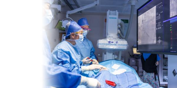
Advances in imaging lead to innovation, new treatment options.
Deepu Nair, MD, FACC
Deepu Nair, MD, FACC
請點擊此轉換成中文
Since the first electrocardiogram (EKG or ECG) was used to measure heartbeats in 1887, diagnosing and treating heart conditions has been through many revolutionary changes. Today, cardiologists at El Camino Health can view the heart and vascular system in a variety of ways, observe how it functions in real time and even perform complex procedures without the need for open-heart surgery.
As a cardiologist and medical director of echocardiography at El Camino Health, I am fortunate to see firsthand how advances in imaging technology can lead not only to easier, more precise diagnosis but also to new, less invasive procedures — and ultimately better care.
New, more advanced imaging technology has dramatically changed cardiovascular care in recent years, and it's still evolving rapidly. At El Camino Health, we're not just keeping up; we're on the front line, advancing diagnosis, treatment and personalized heart care. Honestly it seems like we are constantly adapting our techniques to some new idea, and because we're here in Silicon Valley, we are often among the first to see some developments.
The two biggest game changers in cardiac imaging in recent years have been computed tomography angiography (CTA) and high-resolution, three-dimensional (3D) echocardiography. Though CT scans and echocardiograms aren't new, the technology in both cases has become much more sophisticated — and powerful.
A New Kind of CT Scan
Computed tomography, often called a CT scan or CAT scan, uses X-rays to create detailed images of areas inside the body. CT angiography, one of the game changers I've already mentioned, carries CT scanning much further, providing detailed pictures of the heart and blood vessels throughout the body.
CTA can identify narrow or blocked areas of a blood vessel, as well as the presence of arterial plaque, the fatty substance made of cholesterol and other materials from the blood stream. Being able to detect the presence of plaque is a huge advancement in diagnosis, because plaque is a bigger predictor of long-term heart attack risk than blood pressure, cholesterol levels or other traditional risk factors, so detecting it early can potentially be lifesaving.
This noninvasive test takes less than 10 minutes and presents modest radiation exposure, so it's quite safe. It allows us to get a complete picture of heart arteries. In fact, in many cases, we can use it to rule out coronary disease, which helps the patient avoid unnecessary procedures. If a patient has symptoms for heart disease, this can be a very useful test.
Echocardiography Shows the Heart Working in Real Time
Echocardiography uses ultrasound to monitor heart function and has been in use for decades. But today's high-resolution, three-dimensional, real-time imaging of anatomy and function is not your grandfather's echocardiogram.
The development of high-resolution echo technology with 3D capability is another gamechanger. It allows us to visualize both anatomy and function of the heart. We can see the heart pumping, the valves in motion and other details so we can be very precise about what the problem is. It also helps us determine whether there are minimally invasive or surgical options that would solve the problem.
Better Technology Leads to Innovation: Structural Heart
As imaging technology has improved, research into new, less invasive or more effective treatment options has picked up speed. Being able to see the heart and vascular system in such detail sparks ideas and innovation.
Imaging to guide catheter-based procedures is one of my clinical interests, and what's happening today is very exciting. We're learning how to fix problems that previously had no good solution, and we're developing less invasive procedures for problems that once required open-heart surgery. Many of these less invasive treatment options are in the emerging area of structural heart disease, focusing on anatomical abnormalities of the heart itself, including valves, walls or chambers.
Two examples of recent innovation are the MitraClip and the Watchman device.
The MitraClip, now in use worldwide, was invented by El Camino Health's own Fred St. Goar, MD. El Camino Health's cardiology team was one of the first in California to use the MitraClip to treat mitral valve disease (also called mitral valve regurgitation) — a condition in which the heart valve doesn't close properly. The standard method of treatment is open-heart surgery, which isn’t feasible for many patients because of age or overall health.
The MitraClip is placed using a catheter, a very thin tube, which is passed through a vein in the groin. Advanced imaging guides the placement in the heart and allows function to be assessed with the clip in place so that adjustments can be made right away. This device offers a treatment option for people who have severe symptoms and who are at too great a risk to have traditional surgery.
The Watchman device is a small implant placed in the heart that can reduce the risk of stroke in patients with atrial fibrillation (often called AFib). The balloon-like implant is inserted near the groin and placed inside the left atrial appendage of the heart to prevent blood from pooling there. With this device, we are often able to reduce the need for blood thinners for AFib, while also reducing stroke risk.
Then and Now
Catheter-based (also called percutaneous) treatment options are evolving all the time. I started at El Camino Health in 2008, and since then many of the procedures that were strictly surgical can now also be done percutaneously.
These treatment options are less invasive, with tiny incisions, and may be lower risk than open surgery for some patients. Recovery is often much quicker. Patients can often go home after one or two days.
However, catheter-based solutions aren’t for everyone. Surgical methods are tried and true, and we don’t yet have long-term results from some catheter-based procedures.
Age, overall health, life expectancy, the patient's goals and preferences, and other factors must be considered when making the decision. For instance, in a younger patient, surgery may be the better choice for long-term outcomes, while an older patient is usually at higher risk of surgical complications.
Our team members work closely with patients and families so they can make informed decisions about their care. Everyone is unique, and every case is different. As we learn more, we'll continue to develop newer, safer and more reliable treatment options. Cardiology will always be rapidly evolving as new knowledge is applied, and El Camino Health will continue to be a leader in advancing heart care and saving lives.
Learn more about Heart and Vascular Care at El Camino Health.
This article first appeared in the February 2023 edition of the HealthPerks newsletter.

